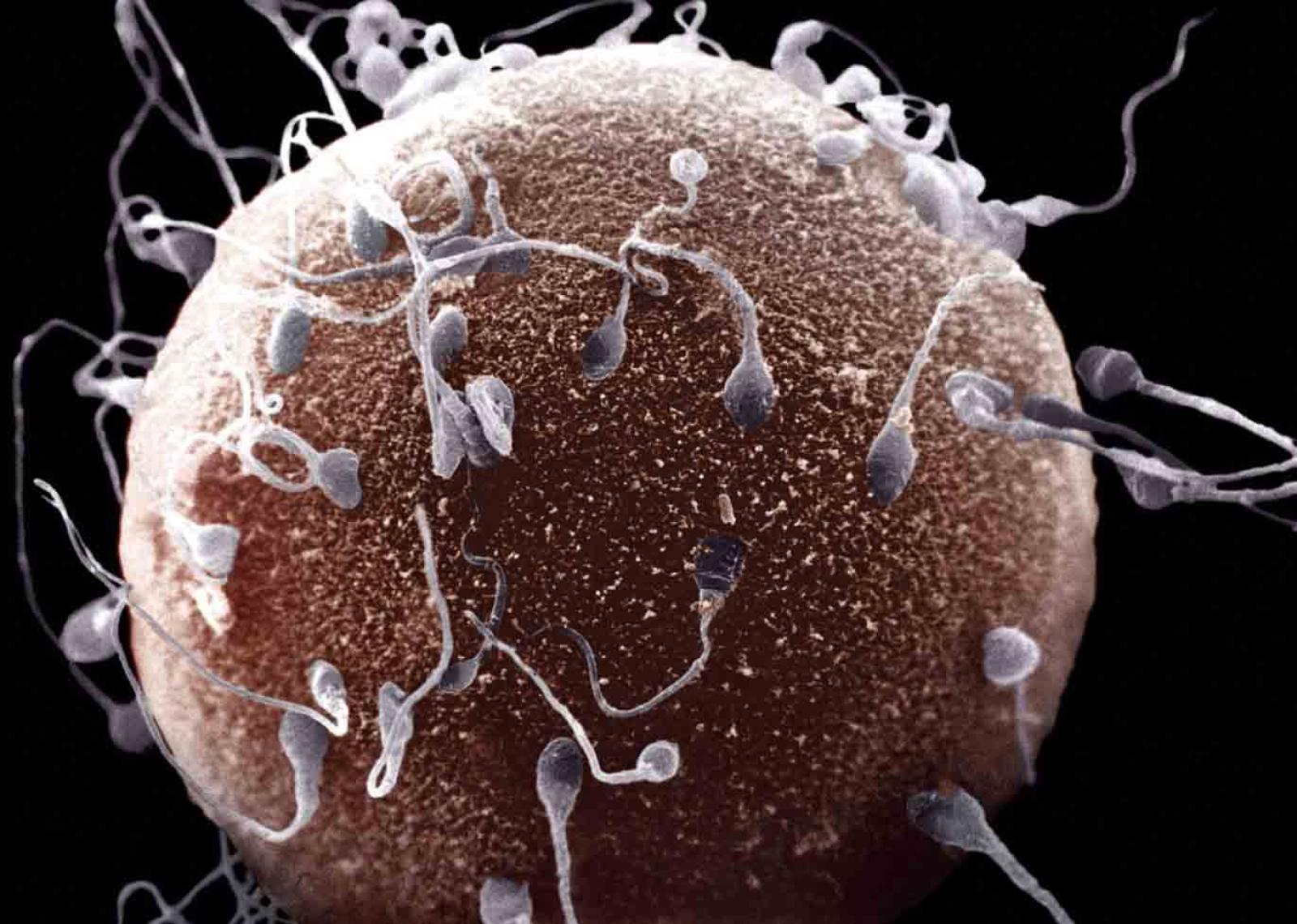
Training a machine to classify sperms based on their physical traits: this is the task accomplished by a group of researchers from the Center for Complexity and Biosystems of the University of Milan, who just published the study on Scientific Report. A task that might be of great help in the field of reproductive medicine.
The presence of abnormalities, such as a large or misshapen head or a crooked or double tail, might affect the ability of the sperm to reach and penetrate an egg. For this reason, sperm morphology is one of the factors that are examined as part of a semen analysis to evaluate male infertility. And such evaluation is usually performed by experts with a trained eye, who look at the semen under a microscope and classify sperms based on their aspect. However, due to the increasing amount of available digital images, it is becoming important to develop automatic techniques of classification and diagnosis.
To do such a thing, it is then necessary to develop reliable automated methods for cell morphology assessment but, at the moment, objective tools exist for sperm motility assessment, while current automatic methods for sperm morphology are still not accurate and difficult to use. Hence, subjective morphology sperm cell assessment is still the standard in laboratories but results in large variability in the outcome.
“Machine learning-based intelligent systems could play a pivotal role to reach this goal”, says the biologist Caterina La Porta, from the Department of Environmental Sciences and Policy, who coordinated the research. “These systems can train themselves to learn the patterns of the data we provide them and produce a prediction model. The final goal is then to be able to automatically classify a data set with unknown labels”.
The researchers focused their attention on the morphology of the acrosome, an organelle with the shape of a head-cap that covers the sperm nucleus. The acrosome contains the enzymes necessary to break down the outer membrane of the egg, allowing the sperm to get inside it and begin the fertilization process. They used a large amount of images of mouse sperms to perform a three-dimensional digital reconstruction of their acrosomes, and then compute a series of parameters such as volume, surface and local curvatures. Finally, they analysed these traits by machine learning and compared them with the ground truth provided by a direct assessment by eye. The algorithm spotted differences that an expert eye was not able to distinguish, and its classifications were corrected in 73% of trials – a higher percentage than those obtained with other methods.
“We have proposed a general strategy to classify acrosomes during the course of sperms development, according to their morphological features”, concludes La Porta. “This could help solve the relevant clinical issue of quantifying the percentage of sperm cells with normal acrosome and therefore assess fertility”.