
Is there life on Mars?
It is five years since the NASA rovers named "Spirit" and "Opportunity" have been exploring Mars, sending their close up pictures and especially the microscope images.(Squyre 2004).
It is five years since the NASA rovers named “Spirit” and “Opportunity” have been exploring Mars, sending their close up pictures and especially the microscope images.(Squyre 2004). The images (on the website http://marsrovers.jpl.nasa.gov/gallery) when studied at high magnification (250-400%) are a good source of information and capture one’s attention for days on end. Often, when looking at them a second time one discovers things that were missed the first time.
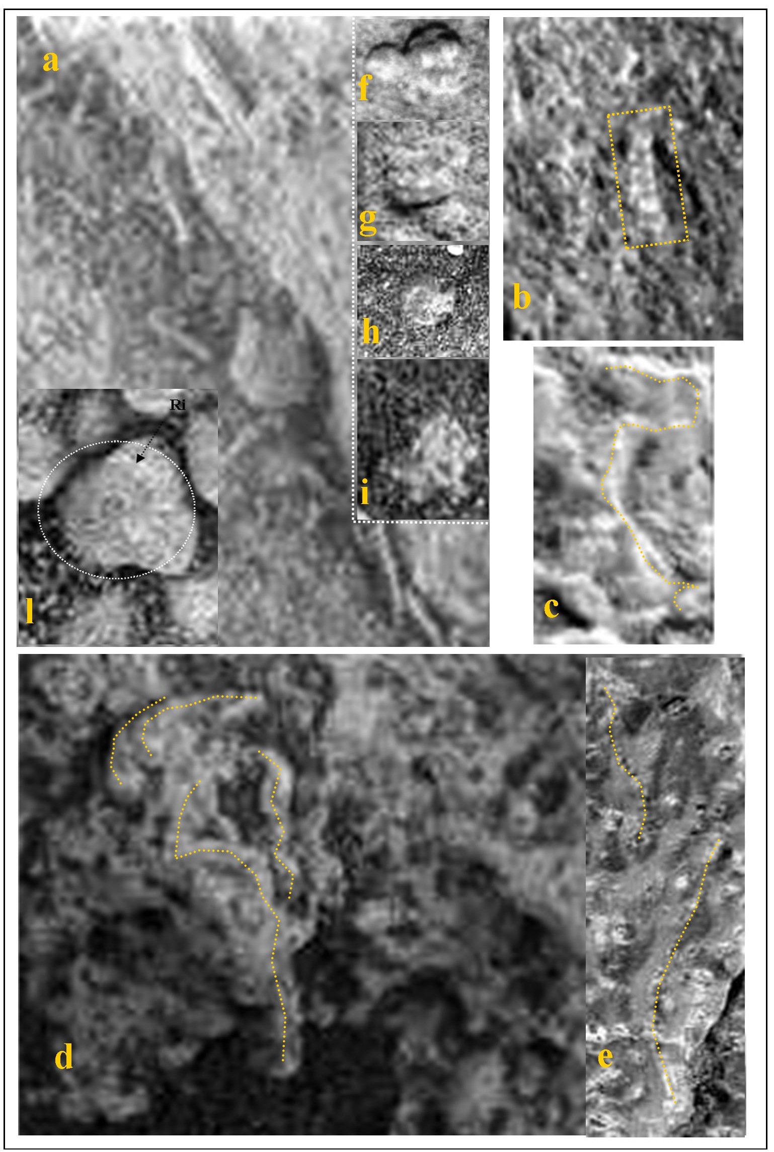
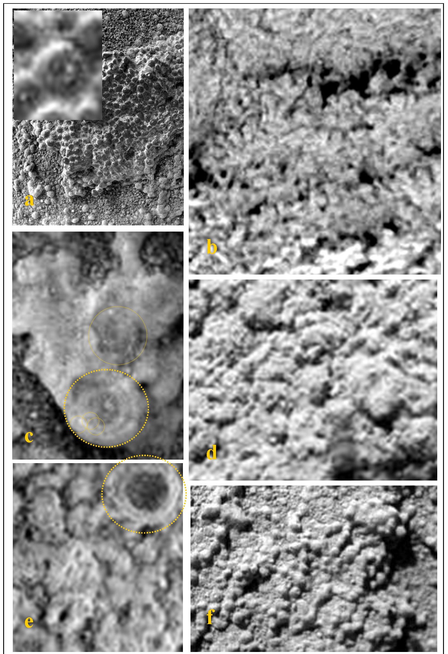
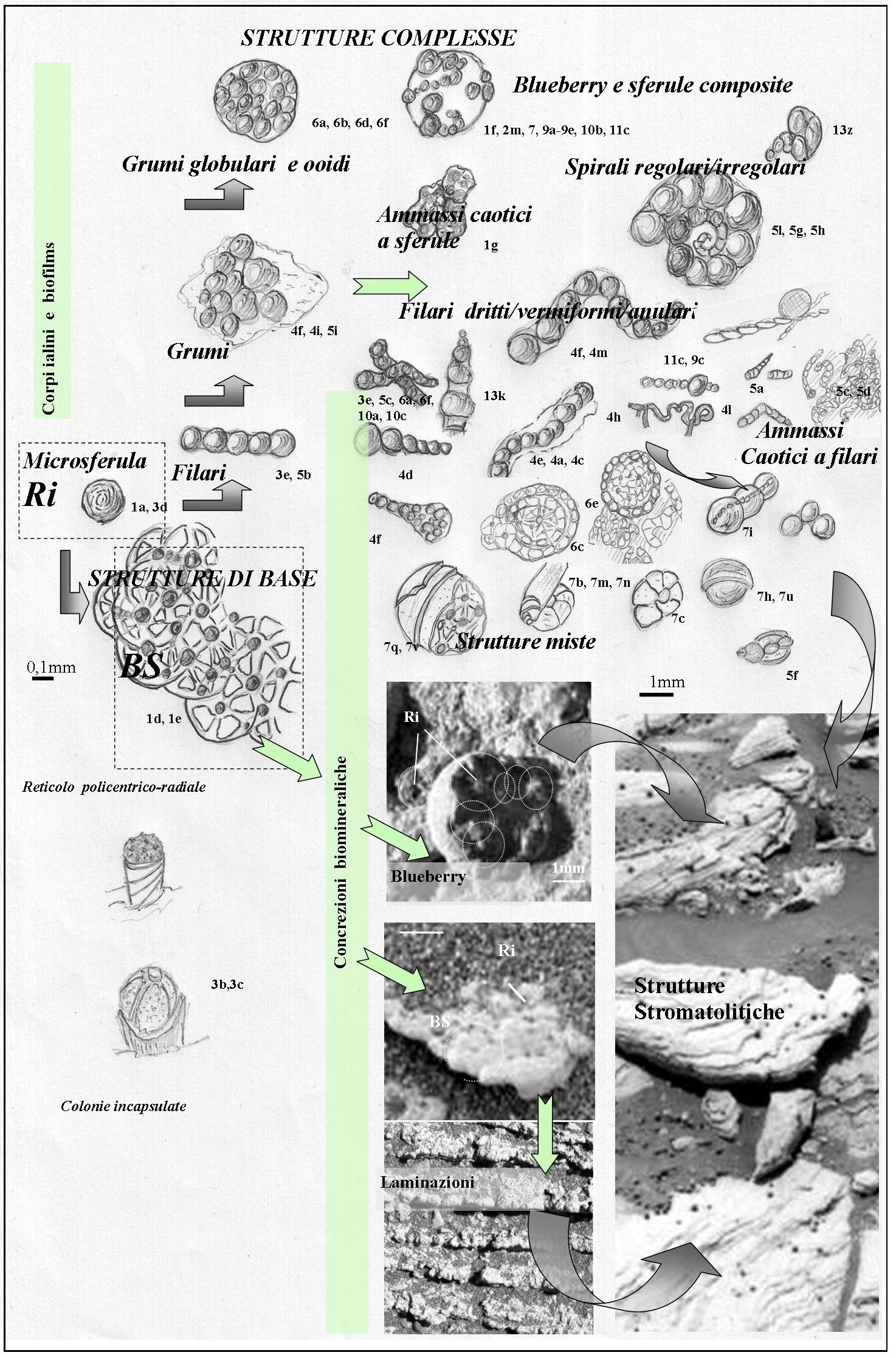
By carefully analyzing the laminated sediments of “Meridiani Planum” of Mars and the strange hematite spherules, known as “blueberries” (Moore 2004), whose images were taken by the rover “Opportunity”, the authors suggested that these are organo-sedimentary structures, that is structures produced by micro-organisms and having, though with all the peculiarities of the Martian world, several common features - on a different scale of observation - with the familiar terrestrial stromatolites (Rizzo and Cantasano 2009; #FFF# figures [1](/files/brilliant_gallery/marte/marte_fig1.jpg “Structure of organo-sedimentary deposits of Mars (the white bar represents 1 mm). In a: hyaline films structured as networks of interconnected “Ri microspherules”, forming a transparent film covering the bodies underneath. These films (in b) trap grains (agglutinated lamina LB), or tend to become opalescent (in c) due to accretion (skeletal lamina LA), forming (d) structures of the “BS” type (the sketch shows one of these structures) which make up the laminated sediments. Consequently, the skeletal laminae have crenellated/segmented edges corresponding to the sides of BS superimposed on flat tile (see boxes). All sedimentary bodies observed at high magnification (mostly serpentine arrays, blueberries and various clusters; in f-l ), are due to the coalescence and biomineralization of «RI» spherules in various spatial configurations.”), [2](/files/brilliant_gallery/marte/marte_Fig2.jpg “Similarities between structures in metre (in a-c) and centimetre / sub-centimetre (in d-n) size, (the white bar represents one centimetre) of organo-sedimentary terrestrial deposits (black label) and similar structures of Martian sediments (yellow label). In a: the typical stromatolite regressive sequence, with a gradual shift from a lower predominantly detrital layer to a mainly biological upper level. In b: polygonal structure. In c: columnar structure. In d: mm lamination characterized by alternation of skeletal (coalescing calcimicrobes) and agglutinated (trapped grains ) layers. In e: rapid transition from flat to curved laminae (one of the features for the recognition of stromatolites). In f: convoluted laminae structure. In g: dendritic structure. In h: thrombolite tree structure. In i: tuff structure In l: oncolite clusters. In m: similarity between terrestrial oncolite and “blueberries” (note the common diametrical features). In n: the presence of a gelatinous material. The images of a, b, c refer to Australian stromatolites, those in d, e, f, are taken from Schopf (Schopf, 1967), those in g, h, i, l, n are taken from Riding (Riding, 2000, 2002).”), [3](/files/brilliant_gallery/marte/marte_Fig3.jpg “Affinity between microscopic structures (yellow label for Martians deposits: the white bar represents 1 mm). In a: thin stromatolite sections showing structures of “BS " type (circles). In the box: a stromatolite showing similar structures at the macroscopic scale. In b: colonies of cyanobacteria, typically encapsulated (arrows). In c: other bacterial colonies of stromatolites. In d: microbial spherical colonies, reproduced in the laboratory having a capacity of orientation and biomineralization, creating ooids of size similar to ‘“RI” microspherules (calcimicrobes; by Brehem et al., 2004). In e: similar “Ri” type structures can be observed both on terrestrial stromatolites (translucent spheres, from Riding, 2000) and on Martian sediments. The latter show interconnected microspherules (examples in boxes), forming linear and serpentine structures (see also figures 4, 5 and 6).”)).These sediments are characterized by a sequential succession of a pair of sub-millimetre laminae, with a whitish concretion layer (purely skeletal / LA) and a silty fine-grained layer (purely agglutinated / LB), embedding several clusters of microspherules ( calcimicrobes), in linear, serpentine or spheroidal arrays, including the “blueberries” themselves (#FFF# figure [3](/files/brilliant_gallery/marte/marte_Fig3.jpg “Affinity between microscopic structures (yellow label for Martians deposits: the white bar represents 1 mm). In a: thin stromatolite sections showing structures of “BS " type (circles). In the box: a stromatolite showing similar structures at the macroscopic scale. In b: colonies of cyanobacteria, typically encapsulated (arrows). In c: other bacterial colonies of stromatolites. In d: microbial spherical colonies, reproduced in the laboratory having a capacity of orientation and biomineralization, creating ooids of size similar to ‘“RI” microspherules (calcimicrobes; by Brehem et al., 2004). In e: similar “Ri” type structures can be observed both on terrestrial stromatolites (translucent spheres, from Riding, 2000) and on Martian sediments. The latter show interconnected microspherules (examples in boxes), forming linear and serpentine structures (see also figures 4, 5 and 6).”), [4](/files/brilliant_gallery/marte/marte_Fig4.jpg “Examples of complex structures, formed by the coalescence of ‘“Ri” microspherules (the white bar represents 1 mm; examples in the boxes, where the coloured lines track the development of some of them). In a, b, d, straight arrays with variable size Ri patterns. In b, c, e, g: wavy arrays, perhaps with a slightly differentiated first segment. In e: first stages of accretion around the calcimicrobe. In b: microspherules with hyaline appearance. In c, g: mineralized appearance. In f: sheath and elongated clusters of microspherules, which tend to be iso-oriented. in h-m: serpentine or granular arrays of microspherules, disorderly arranged.”), 5, 6, [7](/files/brilliant_gallery/marte/marte_fig7.jpg “Morphology and structure of “blueberries”. In a: highly variable forms, depending on the internal structure, far from homogeneous. In b: structure with rolled-up laminae and different rotation directions. In c: spiralled structure. In d: partly rolled-up structure. In e: alveolar structure, rolled-up in a spiral. In f, o: microspherules of different sizes, with gradual transitions to blueberries. In f: curious phenomena of iso-orientation. In g: blueberry made up of a set of microspherules. In h, u: bi-spherical blueberries. In i: coaxial coalescing spherules (one array of microspherules is oddly oriented along the axis of coalescence). In l: clusters of microspherules/ equigranular and clustered spherules (contacts are interpenetrating “cauliflower -like”). In m, n: Blueberries showing some polylobate and some spiralling characteristics. In p: blueberries with texture and colour different from surrounding clusters. In q, r: polycentric structure of blueberries (resulting from BS elements, spatially interconnected). In s: the smaller spherules, with asynchronous growth, is emerging from the “mother” spherule. In t: emerging spherule with a deformed cover. In v: spiral rolling-up of a blueberry, with secondary and eccentric growth centres (both highlighted by circles).”), 8). Sediment that NASA has already attributed to the presence of water in the past (Squyres 2006; Wacey 2009; Jolliff 2006).
The genetic hypothesis relating to microbial activity is consistent with other scientific findings on Mars (Mackay 1996, Mackay 1998, Schulze-Mazuk 2008), where the presence of methane and formaldehyde was reported (Onstott 2006), where a circadian activity, detected in Vicking experiments, is suspected (Van Dongen 2009); while it seems that the existence of “original” nanobacteria fossils in meteorites of Martian origin has recently been established by NASA(news release).
But what’s so peculiar in Mars sediments and where does the microbial hypothesis come from?
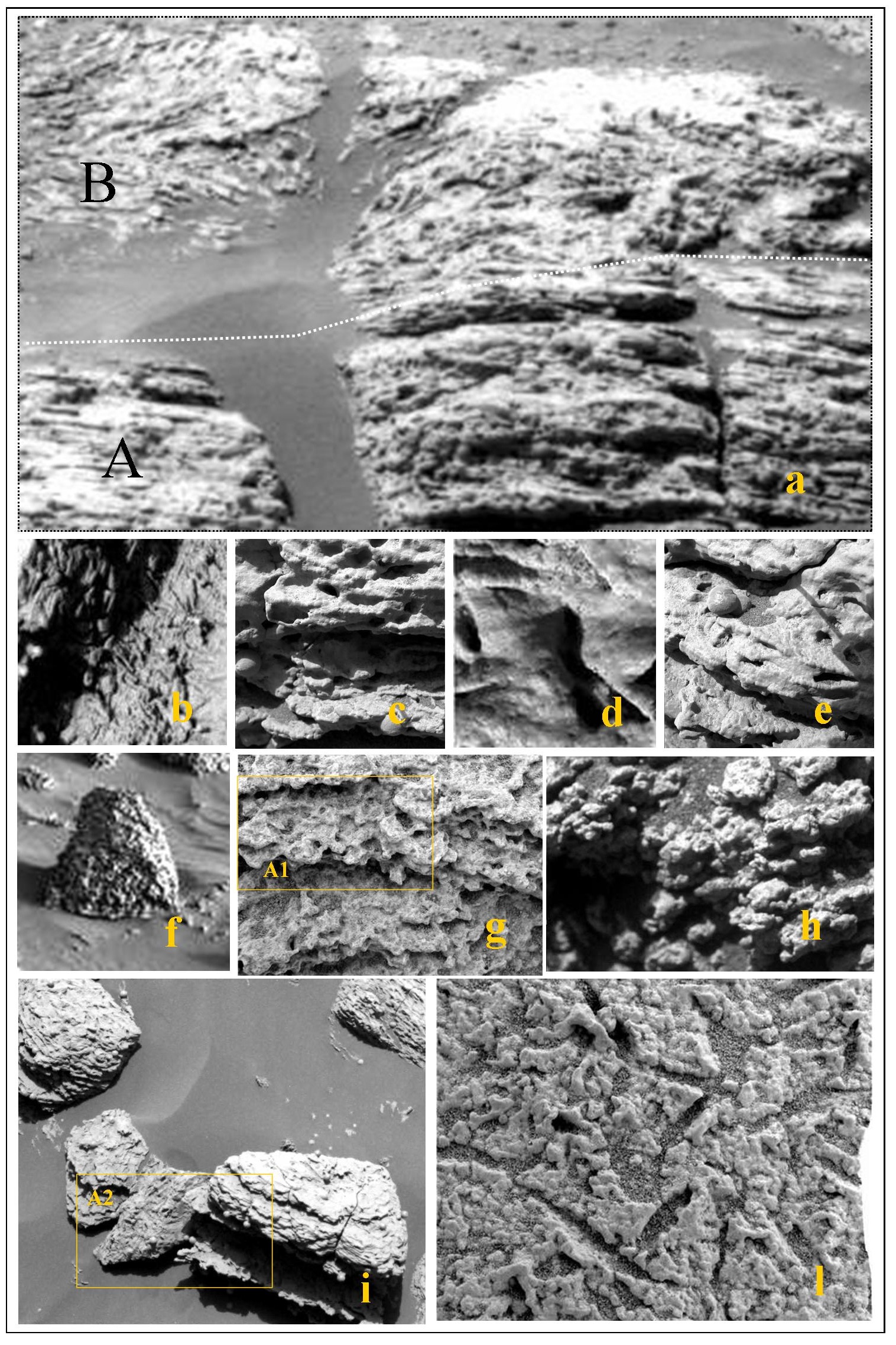
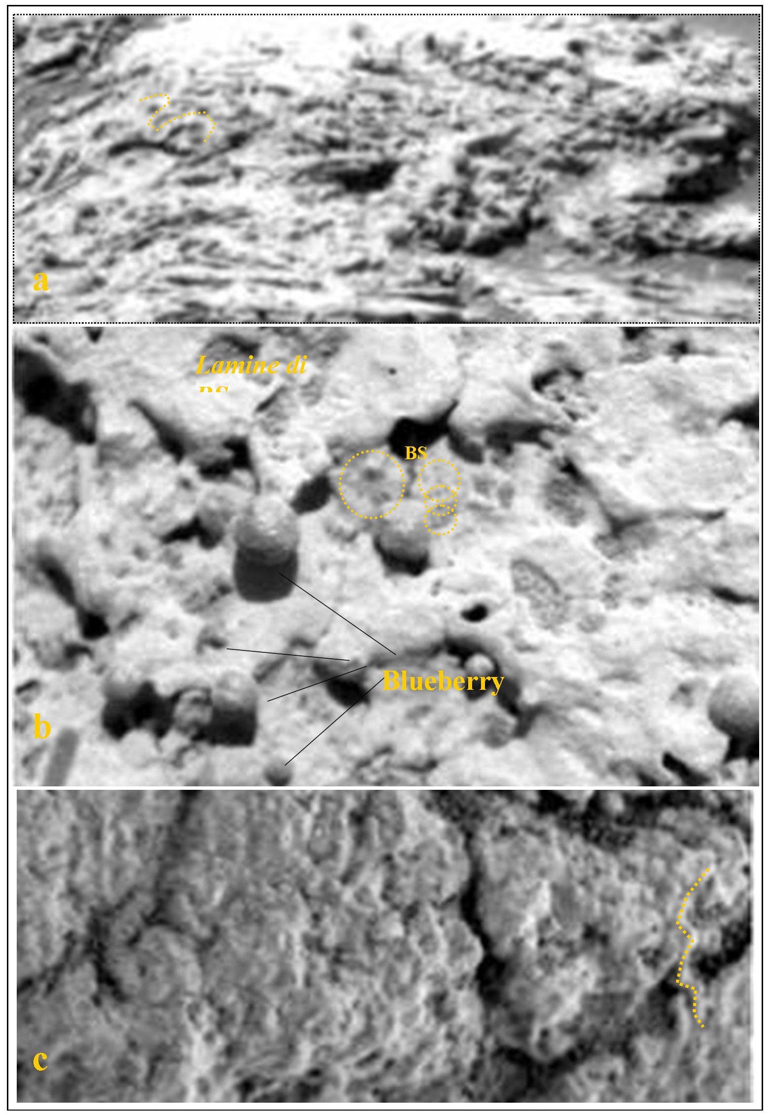
The thin lamination, as it is known, is typical of lake sediments or deposition in calm seas. We note, however, that often the laminated structures are gradually replaced by extremely disordered arrangements, with laminar convolutions filled with cavities in which coalescing serpentine forms develop, whose global texture is similar to the typical characteristics of organogenic environments (#FFF# figures 8, 9, 10). It is often observed that the laminae themselves, in the vicinity of blueberries, tend to encircle them, by changing the plane-parallel structure (#FFF# figure 9). These arrays can be observed in the shots taken by both Opportunity and Spirit (Squyre 2006).

With reference to the textural appearance of laminated sediments it can be observed that LA and LB laminae show similar and repeated patterns (#FFF# figure [1](/files/brilliant_gallery/marte/marte_fig1.jpg “Structure of organo-sedimentary deposits of Mars (the white bar represents 1 mm). In a: hyaline films structured as networks of interconnected “Ri microspherules”, forming a transparent film covering the bodies underneath. These films (in b) trap grains (agglutinated lamina LB), or tend to become opalescent (in c) due to accretion (skeletal lamina LA), forming (d) structures of the “BS” type (the sketch shows one of these structures) which make up the laminated sediments. Consequently, the skeletal laminae have crenellated/segmented edges corresponding to the sides of BS superimposed on flat tile (see boxes). All sedimentary bodies observed at high magnification (mostly serpentine arrays, blueberries and various clusters; in f-l ), are due to the coalescence and biomineralization of «RI» spherules in various spatial configurations.")a-d), (composed of very small spherules (Ri), with a radial array of stable connections, resulting in a “brick-like” base (BS). BS in LB resembles a wheel with cavities interposed between radial connections, while the concretional film in LA gives BS the appearance of a flower and the laminae show crenellated edges accordingly(#FFF# figure [1](/files/brilliant_gallery/marte/marte_fig1.jpg “Structure of organo-sedimentary deposits of Mars (the white bar represents 1 mm). In a: hyaline films structured as networks of interconnected “Ri microspherules”, forming a transparent film covering the bodies underneath. These films (in b) trap grains (agglutinated lamina LB), or tend to become opalescent (in c) due to accretion (skeletal lamina LA), forming (d) structures of the “BS” type (the sketch shows one of these structures) which make up the laminated sediments. Consequently, the skeletal laminae have crenellated/segmented edges corresponding to the sides of BS superimposed on flat tile (see boxes). All sedimentary bodies observed at high magnification (mostly serpentine arrays, blueberries and various clusters; in f-l ), are due to the coalescence and biomineralization of «RI» spherules in various spatial configurations.")e).
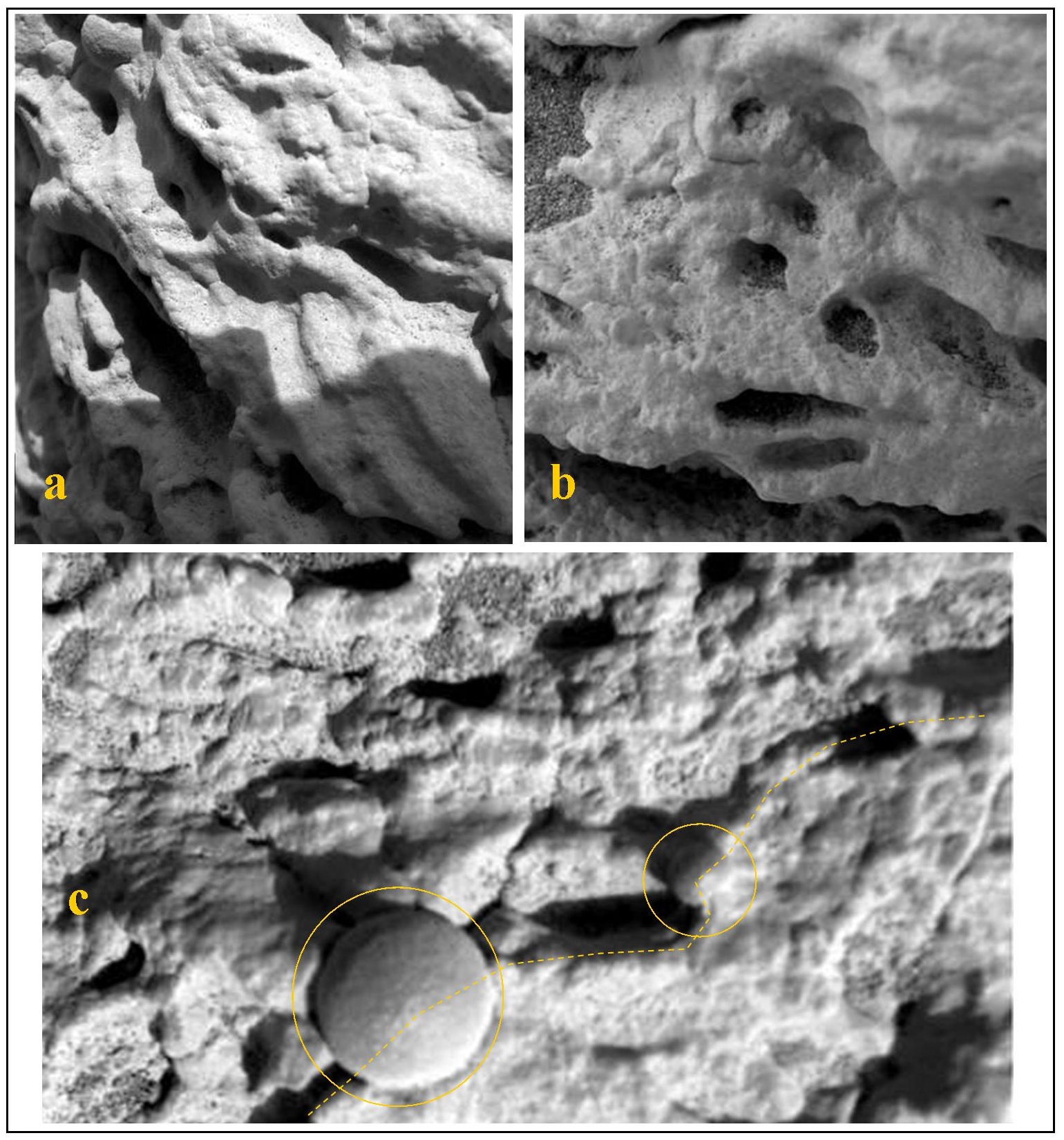
In some cases there is a hyaline veil that has similar patterns, showing progressive accretional processes (biomineralization), with transition from transparent hyaline forms to milky-white coloured forms; this accretion is spread randomly on the exposed surfaces; however, in some cases this accretion clearly departs from the basic or complex identified structures (#FFF# figure [4](/files/brilliant_gallery/marte/marte_Fig4.jpg “Examples of complex structures, formed by the coalescence of ‘“Ri” microspherules (the white bar represents 1 mm; examples in the boxes, where the coloured lines track the development of some of them). In a, b, d, straight arrays with variable size Ri patterns. In b, c, e, g: wavy arrays, perhaps with a slightly differentiated first segment. In e: first stages of accretion around the calcimicrobe. In b: microspherules with hyaline appearance. In c, g: mineralized appearance. In f: sheath and elongated clusters of microspherules, which tend to be iso-oriented. in h-m: serpentine or granular arrays of microspherules, disorderly arranged.")e-f, 5c-e, 5h-i). On the same hyaline film - at times - some scattered granules can be observed, perhaps indicating the beginning of an agglutination process (collagen? #FFF# figure [1](/files/brilliant_gallery/marte/marte_fig1.jpg “Structure of organo-sedimentary deposits of Mars (the white bar represents 1 mm). In a: hyaline films structured as networks of interconnected “Ri microspherules”, forming a transparent film covering the bodies underneath. These films (in b) trap grains (agglutinated lamina LB), or tend to become opalescent (in c) due to accretion (skeletal lamina LA), forming (d) structures of the “BS” type (the sketch shows one of these structures) which make up the laminated sediments. Consequently, the skeletal laminae have crenellated/segmented edges corresponding to the sides of BS superimposed on flat tile (see boxes). All sedimentary bodies observed at high magnification (mostly serpentine arrays, blueberries and various clusters; in f-l ), are due to the coalescence and biomineralization of «RI» spherules in various spatial configurations.")b). The size and the structural arrangements of LA, LB and the hyaline films are definitely similar (#FFF# figure [1](/files/brilliant_gallery/marte/marte_fig1.jpg “Structure of organo-sedimentary deposits of Mars (the white bar represents 1 mm). In a: hyaline films structured as networks of interconnected “Ri microspherules”, forming a transparent film covering the bodies underneath. These films (in b) trap grains (agglutinated lamina LB), or tend to become opalescent (in c) due to accretion (skeletal lamina LA), forming (d) structures of the “BS” type (the sketch shows one of these structures) which make up the laminated sediments. Consequently, the skeletal laminae have crenellated/segmented edges corresponding to the sides of BS superimposed on flat tile (see boxes). All sedimentary bodies observed at high magnification (mostly serpentine arrays, blueberries and various clusters; in f-l ), are due to the coalescence and biomineralization of «RI» spherules in various spatial configurations.”)).
This is, in itself, another anomaly, since from bodies of different composition and in an environment of simple chemico-physical origins one would expect different structures. In addition it is not clear why the accretion phenomena should be more intense around the structures we identified (biogenic origin).
In some cases a skeleton lamina is barely visible, without interposed agglutinated laminae, both because the latter are not very thick and because there is a white film on the surface (with the same aspect of LA), covering the laminated sequence.
At times this film, which in some cases seems to have the density of plastic, is lifted making room to an emerging blueberry , which in such manner carries its remainders upward (#FFF# figure 9a-e). The texture of materials and the processes of “internal” growth (see also #FFF# figure [7](/files/brilliant_gallery/marte/marte_fig7.jpg “Morphology and structure of “blueberries”. In a: highly variable forms, depending on the internal structure, far from homogeneous. In b: structure with rolled-up laminae and different rotation directions. In c: spiralled structure. In d: partly rolled-up structure. In e: alveolar structure, rolled-up in a spiral. In f, o: microspherules of different sizes, with gradual transitions to blueberries. In f: curious phenomena of iso-orientation. In g: blueberry made up of a set of microspherules. In h, u: bi-spherical blueberries. In i: coaxial coalescing spherules (one array of microspherules is oddly oriented along the axis of coalescence). In l: clusters of microspherules/ equigranular and clustered spherules (contacts are interpenetrating “cauliflower -like”). In m, n: Blueberries showing some polylobate and some spiralling characteristics. In p: blueberries with texture and colour different from surrounding clusters. In q, r: polycentric structure of blueberries (resulting from BS elements, spatially interconnected). In s: the smaller spherules, with asynchronous growth, is emerging from the “mother” spherule. In t: emerging spherule with a deformed cover. In v: spiral rolling-up of a blueberry, with secondary and eccentric growth centres (both highlighted by circles).")s) are inconsistent with the mineral world.
The similarity between the Martian structures / textures and those of stromatolites and, more generally, of terrestrial organo-sedimentary sediments, covers several aspects: aspects that develop at different scales, from the minimum scale of observation up to the macroscopic scale of outcrops (#FFF# figure [2](/files/brilliant_gallery/marte/marte_Fig2.jpg “Similarities between structures in metre (in a-c) and centimetre / sub-centimetre (in d-n) size, (the white bar represents one centimetre) of organo-sedimentary terrestrial deposits (black label) and similar structures of Martian sediments (yellow label). In a: the typical stromatolite regressive sequence, with a gradual shift from a lower predominantly detrital layer to a mainly biological upper level. In b: polygonal structure. In c: columnar structure. In d: mm lamination characterized by alternation of skeletal (coalescing calcimicrobes) and agglutinated (trapped grains ) layers. In e: rapid transition from flat to curved laminae (one of the features for the recognition of stromatolites). In f: convoluted laminae structure. In g: dendritic structure. In h: thrombolite tree structure. In i: tuff structure In l: oncolite clusters. In m: similarity between terrestrial oncolite and “blueberries” (note the common diametrical features). In n: the presence of a gelatinous material. The images of a, b, c refer to Australian stromatolites, those in d, e, f, are taken from Schopf (Schopf, 1967), those in g, h, i, l, n are taken from Riding (Riding, 2000, 2002).”), [3](/files/brilliant_gallery/marte/marte_Fig3.jpg “Affinity between microscopic structures (yellow label for Martians deposits: the white bar represents 1 mm). In a: thin stromatolite sections showing structures of “BS " type (circles). In the box: a stromatolite showing similar structures at the macroscopic scale. In b: colonies of cyanobacteria, typically encapsulated (arrows). In c: other bacterial colonies of stromatolites. In d: microbial spherical colonies, reproduced in the laboratory having a capacity of orientation and biomineralization, creating ooids of size similar to ‘“RI” microspherules (calcimicrobes; by Brehem et al., 2004). In e: similar “Ri” type structures can be observed both on terrestrial stromatolites (translucent spheres, from Riding, 2000) and on Martian sediments. The latter show interconnected microspherules (examples in boxes), forming linear and serpentine structures (see also figures 4, 5 and 6).”)). Generally, random morphological similarity between different bodies is a sporadic fact and a coincidence; it is hard to imagine such a coincidence repeated at different scales, which is more inherent in the internal structure of bodies rather than in their simple morphology
But what is most interesting is that almost all sedimentary bodies observed at sub-millimetre scale, while having different resulting forms are made up of coalescing Ri microspherules , whose different spatial organization (in lines, planes or clusters; according to regular or disordered geometries ) gives rise to more complex structures.
The size of the Ri microspherules are within the limit of resolution, equal to 0.04 mm up to 0.3 mm, while the basic structures of laminated bodies BS and Ri blueberries themselves are about 0.1 mm (#FFF# figure [1](/files/brilliant_gallery/marte/marte_fig1.jpg “Structure of organo-sedimentary deposits of Mars (the white bar represents 1 mm). In a: hyaline films structured as networks of interconnected “Ri microspherules”, forming a transparent film covering the bodies underneath. These films (in b) trap grains (agglutinated lamina LB), or tend to become opalescent (in c) due to accretion (skeletal lamina LA), forming (d) structures of the “BS” type (the sketch shows one of these structures) which make up the laminated sediments. Consequently, the skeletal laminae have crenellated/segmented edges corresponding to the sides of BS superimposed on flat tile (see boxes). All sedimentary bodies observed at high magnification (mostly serpentine arrays, blueberries and various clusters; in f-l ), are due to the coalescence and biomineralization of «RI» spherules in various spatial configurations.”)).
Therefore it is possible to observe, within the size range of Ri and up to a few millimetres, some rather bizarre products of coalescence, such as (#FFF# figure [1](/files/brilliant_gallery/marte/marte_fig1.jpg “Structure of organo-sedimentary deposits of Mars (the white bar represents 1 mm). In a: hyaline films structured as networks of interconnected “Ri microspherules”, forming a transparent film covering the bodies underneath. These films (in b) trap grains (agglutinated lamina LB), or tend to become opalescent (in c) due to accretion (skeletal lamina LA), forming (d) structures of the “BS” type (the sketch shows one of these structures) which make up the laminated sediments. Consequently, the skeletal laminae have crenellated/segmented edges corresponding to the sides of BS superimposed on flat tile (see boxes). All sedimentary bodies observed at high magnification (mostly serpentine arrays, blueberries and various clusters; in f-l ), are due to the coalescence and biomineralization of «RI» spherules in various spatial configurations.”), [2](/files/brilliant_gallery/marte/marte_Fig2.jpg “Similarities between structures in metre (in a-c) and centimetre / sub-centimetre (in d-n) size, (the white bar represents one centimetre) of organo-sedimentary terrestrial deposits (black label) and similar structures of Martian sediments (yellow label). In a: the typical stromatolite regressive sequence, with a gradual shift from a lower predominantly detrital layer to a mainly biological upper level. In b: polygonal structure. In c: columnar structure. In d: mm lamination characterized by alternation of skeletal (coalescing calcimicrobes) and agglutinated (trapped grains ) layers. In e: rapid transition from flat to curved laminae (one of the features for the recognition of stromatolites). In f: convoluted laminae structure. In g: dendritic structure. In h: thrombolite tree structure. In i: tuff structure In l: oncolite clusters. In m: similarity between terrestrial oncolite and “blueberries” (note the common diametrical features). In n: the presence of a gelatinous material. The images of a, b, c refer to Australian stromatolites, those in d, e, f, are taken from Schopf (Schopf, 1967), those in g, h, i, l, n are taken from Riding (Riding, 2000, 2002).”), [3](/files/brilliant_gallery/marte/marte_Fig3.jpg “Affinity between microscopic structures (yellow label for Martians deposits: the white bar represents 1 mm). In a: thin stromatolite sections showing structures of “BS " type (circles). In the box: a stromatolite showing similar structures at the macroscopic scale. In b: colonies of cyanobacteria, typically encapsulated (arrows). In c: other bacterial colonies of stromatolites. In d: microbial spherical colonies, reproduced in the laboratory having a capacity of orientation and biomineralization, creating ooids of size similar to ‘“RI” microspherules (calcimicrobes; by Brehem et al., 2004). In e: similar “Ri” type structures can be observed both on terrestrial stromatolites (translucent spheres, from Riding, 2000) and on Martian sediments. The latter show interconnected microspherules (examples in boxes), forming linear and serpentine structures (see also figures 4, 5 and 6).”), [4](/files/brilliant_gallery/marte/marte_Fig4.jpg “Examples of complex structures, formed by the coalescence of ‘“Ri” microspherules (the white bar represents 1 mm; examples in the boxes, where the coloured lines track the development of some of them). In a, b, d, straight arrays with variable size Ri patterns. In b, c, e, g: wavy arrays, perhaps with a slightly differentiated first segment. In e: first stages of accretion around the calcimicrobe. In b: microspherules with hyaline appearance. In c, g: mineralized appearance. In f: sheath and elongated clusters of microspherules, which tend to be iso-oriented. in h-m: serpentine or granular arrays of microspherules, disorderly arranged.”), 5, 6, [7](/files/brilliant_gallery/marte/marte_fig7.jpg “Morphology and structure of “blueberries”. In a: highly variable forms, depending on the internal structure, far from homogeneous. In b: structure with rolled-up laminae and different rotation directions. In c: spiralled structure. In d: partly rolled-up structure. In e: alveolar structure, rolled-up in a spiral. In f, o: microspherules of different sizes, with gradual transitions to blueberries. In f: curious phenomena of iso-orientation. In g: blueberry made up of a set of microspherules. In h, u: bi-spherical blueberries. In i: coaxial coalescing spherules (one array of microspherules is oddly oriented along the axis of coalescence). In l: clusters of microspherules/ equigranular and clustered spherules (contacts are interpenetrating “cauliflower -like”). In m, n: Blueberries showing some polylobate and some spiralling characteristics. In p: blueberries with texture and colour different from surrounding clusters. In q, r: polycentric structure of blueberries (resulting from BS elements, spatially interconnected). In s: the smaller spherules, with asynchronous growth, is emerging from the “mother” spherule. In t: emerging spherule with a deformed cover. In v: spiral rolling-up of a blueberry, with secondary and eccentric growth centres (both highlighted by circles).”), 8, 9, 10, 11, 12):
- • arrays of equidimensional or regularly sized spherules (#FFF# figure [4](/files/brilliant_gallery/marte/marte_Fig4.jpg “Examples of complex structures, formed by the coalescence of ‘“Ri” microspherules (the white bar represents 1 mm; examples in the boxes, where the coloured lines track the development of some of them). In a, b, d, straight arrays with variable size Ri patterns. In b, c, e, g: wavy arrays, perhaps with a slightly differentiated first segment. In e: first stages of accretion around the calcimicrobe. In b: microspherules with hyaline appearance. In c, g: mineralized appearance. In f: sheath and elongated clusters of microspherules, which tend to be iso-oriented. in h-m: serpentine or granular arrays of microspherules, disorderly arranged.”));
- • rolling-up spiralled spherules (#FFF# figure 5);
- • BS imbricate structures (#FFF# figure [1](/files/brilliant_gallery/marte/marte_fig1.jpg “Structure of organo-sedimentary deposits of Mars (the white bar represents 1 mm). In a: hyaline films structured as networks of interconnected “Ri microspherules”, forming a transparent film covering the bodies underneath. These films (in b) trap grains (agglutinated lamina LB), or tend to become opalescent (in c) due to accretion (skeletal lamina LA), forming (d) structures of the “BS” type (the sketch shows one of these structures) which make up the laminated sediments. Consequently, the skeletal laminae have crenellated/segmented edges corresponding to the sides of BS superimposed on flat tile (see boxes). All sedimentary bodies observed at high magnification (mostly serpentine arrays, blueberries and various clusters; in f-l ), are due to the coalescence and biomineralization of «RI» spherules in various spatial configurations.”));
- • arrays of serpentine and / or tubular spherules (#FFF# figure 9), sometimes overlapping (figure [1](/files/brilliant_gallery/marte/marte_fig1.jpg “Structure of organo-sedimentary deposits of Mars (the white bar represents 1 mm). In a: hyaline films structured as networks of interconnected “Ri microspherules”, forming a transparent film covering the bodies underneath. These films (in b) trap grains (agglutinated lamina LB), or tend to become opalescent (in c) due to accretion (skeletal lamina LA), forming (d) structures of the “BS” type (the sketch shows one of these structures) which make up the laminated sediments. Consequently, the skeletal laminae have crenellated/segmented edges corresponding to the sides of BS superimposed on flat tile (see boxes). All sedimentary bodies observed at high magnification (mostly serpentine arrays, blueberries and various clusters; in f-l ), are due to the coalescence and biomineralization of «RI» spherules in various spatial configurations.”), [4](/files/brilliant_gallery/marte/marte_Fig4.jpg “Examples of complex structures, formed by the coalescence of ‘“Ri” microspherules (the white bar represents 1 mm; examples in the boxes, where the coloured lines track the development of some of them). In a, b, d, straight arrays with variable size Ri patterns. In b, c, e, g: wavy arrays, perhaps with a slightly differentiated first segment. In e: first stages of accretion around the calcimicrobe. In b: microspherules with hyaline appearance. In c, g: mineralized appearance. In f: sheath and elongated clusters of microspherules, which tend to be iso-oriented. in h-m: serpentine or granular arrays of microspherules, disorderly arranged.”), 5, 10);
- • sometimes arranged side by side forming regular / irregular dislocations either in closed shape or in bundles (#FFF# figure [2](/files/brilliant_gallery/marte/marte_Fig2.jpg “Similarities between structures in metre (in a-c) and centimetre / sub-centimetre (in d-n) size, (the white bar represents one centimetre) of organo-sedimentary terrestrial deposits (black label) and similar structures of Martian sediments (yellow label). In a: the typical stromatolite regressive sequence, with a gradual shift from a lower predominantly detrital layer to a mainly biological upper level. In b: polygonal structure. In c: columnar structure. In d: mm lamination characterized by alternation of skeletal (coalescing calcimicrobes) and agglutinated (trapped grains ) layers. In e: rapid transition from flat to curved laminae (one of the features for the recognition of stromatolites). In f: convoluted laminae structure. In g: dendritic structure. In h: thrombolite tree structure. In i: tuff structure In l: oncolite clusters. In m: similarity between terrestrial oncolite and “blueberries” (note the common diametrical features). In n: the presence of a gelatinous material. The images of a, b, c refer to Australian stromatolites, those in d, e, f, are taken from Schopf (Schopf, 1967), those in g, h, i, l, n are taken from Riding (Riding, 2000, 2002).”));
- • sudden transitions from straight to curved arrays (#FFF# figure [2](/files/brilliant_gallery/marte/marte_Fig2.jpg “Similarities between structures in metre (in a-c) and centimetre / sub-centimetre (in d-n) size, (the white bar represents one centimetre) of organo-sedimentary terrestrial deposits (black label) and similar structures of Martian sediments (yellow label). In a: the typical stromatolite regressive sequence, with a gradual shift from a lower predominantly detrital layer to a mainly biological upper level. In b: polygonal structure. In c: columnar structure. In d: mm lamination characterized by alternation of skeletal (coalescing calcimicrobes) and agglutinated (trapped grains ) layers. In e: rapid transition from flat to curved laminae (one of the features for the recognition of stromatolites). In f: convoluted laminae structure. In g: dendritic structure. In h: thrombolite tree structure. In i: tuff structure In l: oncolite clusters. In m: similarity between terrestrial oncolite and “blueberries” (note the common diametrical features). In n: the presence of a gelatinous material. The images of a, b, c refer to Australian stromatolites, those in d, e, f, are taken from Schopf (Schopf, 1967), those in g, h, i, l, n are taken from Riding (Riding, 2000, 2002).”));
- • gradual transitions from planar laminae to convoluted laminae/ disordered / thrombolitic / dendritic, clusters with cavities (presumably primary #FFF# figures [2](/files/brilliant_gallery/marte/marte_Fig2.jpg “Similarities between structures in metre (in a-c) and centimetre / sub-centimetre (in d-n) size, (the white bar represents one centimetre) of organo-sedimentary terrestrial deposits (black label) and similar structures of Martian sediments (yellow label). In a: the typical stromatolite regressive sequence, with a gradual shift from a lower predominantly detrital layer to a mainly biological upper level. In b: polygonal structure. In c: columnar structure. In d: mm lamination characterized by alternation of skeletal (coalescing calcimicrobes) and agglutinated (trapped grains ) layers. In e: rapid transition from flat to curved laminae (one of the features for the recognition of stromatolites). In f: convoluted laminae structure. In g: dendritic structure. In h: thrombolite tree structure. In i: tuff structure In l: oncolite clusters. In m: similarity between terrestrial oncolite and “blueberries” (note the common diametrical features). In n: the presence of a gelatinous material. The images of a, b, c refer to Australian stromatolites, those in d, e, f, are taken from Schopf (Schopf, 1967), those in g, h, i, l, n are taken from Riding (Riding, 2000, 2002).”), 8 e 9);
- • clusters of microspherules, making up lumps, sheaths, and larger spherules, including ooids (oolites / oncolites) and the blueberries themselves (#FFF# figure 6 e [7](/files/brilliant_gallery/marte/marte_fig7.jpg “Morphology and structure of “blueberries”. In a: highly variable forms, depending on the internal structure, far from homogeneous. In b: structure with rolled-up laminae and different rotation directions. In c: spiralled structure. In d: partly rolled-up structure. In e: alveolar structure, rolled-up in a spiral. In f, o: microspherules of different sizes, with gradual transitions to blueberries. In f: curious phenomena of iso-orientation. In g: blueberry made up of a set of microspherules. In h, u: bi-spherical blueberries. In i: coaxial coalescing spherules (one array of microspherules is oddly oriented along the axis of coalescence). In l: clusters of microspherules/ equigranular and clustered spherules (contacts are interpenetrating “cauliflower -like”). In m, n: Blueberries showing some polylobate and some spiralling characteristics. In p: blueberries with texture and colour different from surrounding clusters. In q, r: polycentric structure of blueberries (resulting from BS elements, spatially interconnected). In s: the smaller spherules, with asynchronous growth, is emerging from the “mother” spherule. In t: emerging spherule with a deformed cover. In v: spiral rolling-up of a blueberry, with secondary and eccentric growth centres (both highlighted by circles).”)).
The much discussed “blueberries”, in particular, are not homogeneous and have varying shapes and structures (#FFF# figure [7](/files/brilliant_gallery/marte/marte_fig7.jpg “Morphology and structure of “blueberries”. In a: highly variable forms, depending on the internal structure, far from homogeneous. In b: structure with rolled-up laminae and different rotation directions. In c: spiralled structure. In d: partly rolled-up structure. In e: alveolar structure, rolled-up in a spiral. In f, o: microspherules of different sizes, with gradual transitions to blueberries. In f: curious phenomena of iso-orientation. In g: blueberry made up of a set of microspherules. In h, u: bi-spherical blueberries. In i: coaxial coalescing spherules (one array of microspherules is oddly oriented along the axis of coalescence). In l: clusters of microspherules/ equigranular and clustered spherules (contacts are interpenetrating “cauliflower -like”). In m, n: Blueberries showing some polylobate and some spiralling characteristics. In p: blueberries with texture and colour different from surrounding clusters. In q, r: polycentric structure of blueberries (resulting from BS elements, spatially interconnected). In s: the smaller spherules, with asynchronous growth, is emerging from the “mother” spherule. In t: emerging spherule with a deformed cover. In v: spiral rolling-up of a blueberry, with secondary and eccentric growth centres (both highlighted by circles).”)).They show both radial polycentric structures of “interconnected Ri” and poly-spherical structures (microspherules emerging from the surface, side-by-side, intersected or even protruding from the " mother " spherule) and also rolled-up laminated structures (sometimes with variables convolutions within the spherules themselves)
The polycentric and rolled-up laminated structures of “blueberries” can be seen as the result of a different Ri dislocation, where the external appearance and internal structure depends on the predominance of their dimensional asymmetry and spatial interconnection links, and, in fact, some reveal intermediate forms, with combined polyspheric and spiralling characteristics. (#FFF# figure [7](/files/brilliant_gallery/marte/marte_fig7.jpg “Morphology and structure of “blueberries”. In a: highly variable forms, depending on the internal structure, far from homogeneous. In b: structure with rolled-up laminae and different rotation directions. In c: spiralled structure. In d: partly rolled-up structure. In e: alveolar structure, rolled-up in a spiral. In f, o: microspherules of different sizes, with gradual transitions to blueberries. In f: curious phenomena of iso-orientation. In g: blueberry made up of a set of microspherules. In h, u: bi-spherical blueberries. In i: coaxial coalescing spherules (one array of microspherules is oddly oriented along the axis of coalescence). In l: clusters of microspherules/ equigranular and clustered spherules (contacts are interpenetrating “cauliflower -like”). In m, n: Blueberries showing some polylobate and some spiralling characteristics. In p: blueberries with texture and colour different from surrounding clusters. In q, r: polycentric structure of blueberries (resulting from BS elements, spatially interconnected). In s: the smaller spherules, with asynchronous growth, is emerging from the “mother” spherule. In t: emerging spherule with a deformed cover. In v: spiral rolling-up of a blueberry, with secondary and eccentric growth centres (both highlighted by circles).”)).
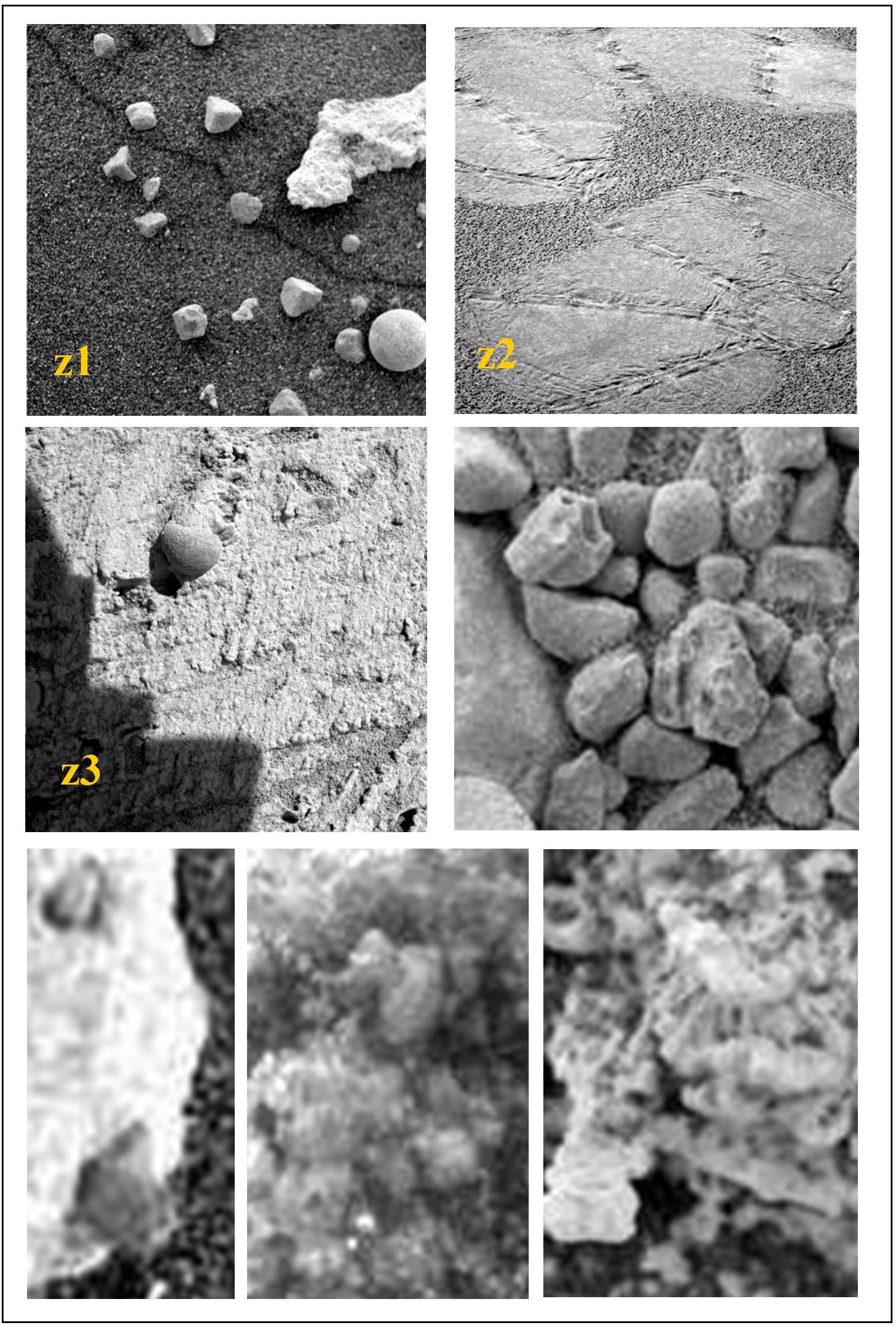
All these structures are quite atypical, and in many respects resemble the structures of carbonate sediments of microbial origin (Riding 2000; Riding 2002) and it is difficult to imagine how they could be the result of a simple physico-chemical process. So that the “trivial microspherules” become “interesting subjects”, due to the nature and complexity of their products, some of which play a “key role” for interpretative purposes. The diversity and variety of forms suggest that they are produced by “builders” smaller than themselves, with coalescence, orientation and accretion features and with repetitive building patterns (in the plane and in the space) at different scales. Moreover, the most basic structures closely resemble those of the bacterial world: the so-called calcimicrobes, which are nothing but coalescing microbial colonies , isorientated and capable of facilitating / triggering concretional processes from the surrounding environment, mostly of spherical or discoid shape(#FFF# figure [3](/files/brilliant_gallery/marte/marte_Fig3.jpg “Affinity between microscopic structures (yellow label for Martians deposits: the white bar represents 1 mm). In a: thin stromatolite sections showing structures of “BS " type (circles). In the box: a stromatolite showing similar structures at the macroscopic scale. In b: colonies of cyanobacteria, typically encapsulated (arrows). In c: other bacterial colonies of stromatolites. In d: microbial spherical colonies, reproduced in the laboratory having a capacity of orientation and biomineralization, creating ooids of size similar to ‘“RI” microspherules (calcimicrobes; by Brehem et al., 2004). In e: similar “Ri” type structures can be observed both on terrestrial stromatolites (translucent spheres, from Riding, 2000) and on Martian sediments. The latter show interconnected microspherules (examples in boxes), forming linear and serpentine structures (see also figures 4, 5 and 6).”)). Recent studies have shown that ooids, whose aggregation forms the well-known terrestrial oolitic concretions, can be obtained in the laboratory and are the result of bacterial activity (Brehemen 2004). Concretions very similar to oolites also exist on Mars, where the internal structure shows arrays consistent with the structures described above (#FFF# figure 6), lacking however the characteristic concentric layers with repeated colours that characterize the terrestrial deposits, due to the recurring climatic changes and, for this reason as well, it is likely that they may be of biogenic origin.
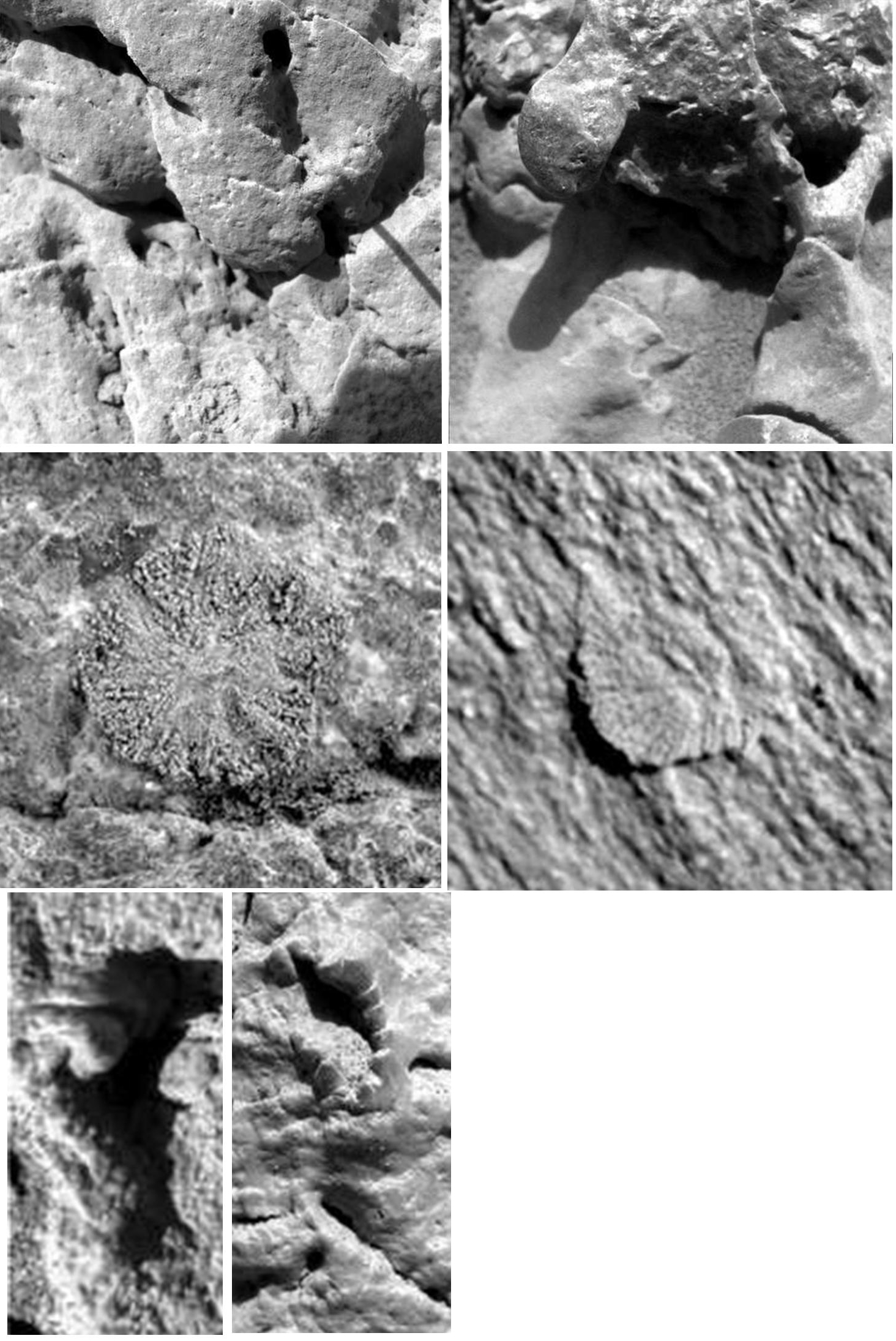
Subject to the assumption on stromatolites , briefly stated in the #FFF# figure 12, the hypothesis that some other selected shapes (#FFF# figures 13, 14 e 15) may represent specific species and other forms of life in fossil state and in part still alive (#FFF# figure 13, image X1) cannot be ruled out. Apart from the limitations to a clear understanding and characterization due to the scantiness of findings, their existence strengthens the hypothesis that, on Mars, other forms of life exist, or at least have existed.

Figures
Brehm U et al. Laboratory cultures of calcifying biomicrospheres generate oids. A contribution to the origin of oolites. Notebooks on Geology, Maintenon, Letters 2004/03 (CG 2004-L03).
Jolliff BL et al. Evidence for Water at Meridiani. Elements 2006; 2 (3): 163.
McKay DS et al. Search for past life on Mars: Possible Relic Biogenic Activity in Martian Meteorite ALH84001. Science 1996; 273: 924.
McKay G et al. Fracture fillings in ALH84001. Feldspatic glass: carbonatic and silica. 29° Annual Lunar and planetary Science Conference 1998, Abstract no. 1944.
Moore JM. Blueberry fields for ever. Nature 2004; 428: 711.
Onstott TC et al. Martian CH4: Sources, Flux and Detection. Astrobiology 2006; 6 (2): 377
Riding, R. (2000). Microbial carbonates: the geological record of calcified bacteria–algal mats and biofilms. Sedimentology 2000; 47 (Suppl. 1): 179.
Riding R. Structure and composition of organic reefs and carbonate mounds: concepts and categories. Earth Science Review 2002; 258: 163.
Rizzo V et al. Possible organosedimentary structures on Mars. International Journal of Astrobiology 2009; 8 (4): 267.
Schulze-Mazuch D et al. The case for life on Mars. International Journal of Astrobiology 2008; 7: 117.
Schopf, J.W. & Barghoorn, E.S. (1967). Alga-like fossils from the Early Precambrian of South Africa. Science 156, 508-512.
Squyres SW et al. The Opportunity Rover’s Athena Science Investigation at Meridiani Planum, Mars. Science 2004; 306: 1698.
Squyres SW et al. Planetary science: bedrock formation at Meridiani Planum. Nature 2006; 443: 1709.
Van Dongen HPA et al. A circadian biosignature in the Labeled Release data from Mars? Proceedings of SPIE 2005; 5906 (59060C): 1.
Wacey D. Early Life on Earth: A Practical Guide. Springer 2009: 1.
Vincenzo Rizzo
Geologia, Università di Firenze, Consiglio Nazionale delle Ricerche
The Scienza in rete newsletter
Every week, free, science picked by hand: research, health, environment. Plus a comment on current events in the editorial by the editor, or by another of us, reserved for newsletter subscribers.